

-
-
 ΠΡΟΪΟΝΤΑ
-
-
-
- Ζέρσεϋ
- Επίδεσμοι δακτύλων
- Ελαστικοί Επίδεσμοι
- Tape συγκράτησηs
- Περιποίηση δέρματος και έλκους
- Εργαλεία μέτρησης και θεραπείας
- Ανομοιόμορφα υποστρώματα
- Αφρώδη υποστρώματα και επιθέματα
- Πακέτα Υλικών Θεραπείας
- Αυτοπερίδεση - Περίδεση ύπνου
- Βοηθήματα εφαρμογής ενδυμάτων
- Ενδύματα διαβαθμισμένης συμπίεσης
- Προστατευτικά μπάνιου
-
-
-
-
-
-
-
-
-
-
-
-
-
-
-
-
-
- Μυοσκελετικά
- Θεραπευτική άσκηση
- Εμβιομηχανική - Κινησιολογία
- Manual Therapy
- Κλινικός συλλογισμός
- Θεραπευτικές τεχνικές
- Trigger Points
- Διαχείριση περιτονίας
- Νευρολογικά
- Taping - Περίδεση
- Ανατομία - Φυσιολογία
- Εναλλακτικές θεραπείες
- Βελονισμός
- Αφίσες ανατομίας
- Λεξικά
- Οίδημα - Λεμφοίδημα
- Ψυχολογία
- Οστεοπαθητική
- Aναπνευστική/Καρδιαγγειακή φυσικοθεραπεία
- Αθλητική φυσικοθεραπεία
- Προσφορές
-
-
-
-
-
-
-
-
-
-
-
-
-
-
-
-
 ESSENTIALS
-
 SUPER OFFERS
-
 ΕΠΙΚΟΙΝΩΝΙΑ
-
 BLOG
BLOG
-
 BRANDS


-
-
 ΠΡΟΪΟΝΤΑ
-
-
-
- Ζέρσεϋ
- Επίδεσμοι δακτύλων
- Ελαστικοί Επίδεσμοι
- Tape συγκράτησηs
- Περιποίηση δέρματος και έλκους
- Εργαλεία μέτρησης και θεραπείας
- Ανομοιόμορφα υποστρώματα
- Αφρώδη υποστρώματα και επιθέματα
- Πακέτα Υλικών Θεραπείας
- Αυτοπερίδεση - Περίδεση ύπνου
- Βοηθήματα εφαρμογής ενδυμάτων
- Ενδύματα διαβαθμισμένης συμπίεσης
- Προστατευτικά μπάνιου
-
-
-
-
-
-
-
-
-
-
-
-
-
-
-
-
-
- Μυοσκελετικά
- Θεραπευτική άσκηση
- Εμβιομηχανική - Κινησιολογία
- Manual Therapy
- Κλινικός συλλογισμός
- Θεραπευτικές τεχνικές
- Trigger Points
- Διαχείριση περιτονίας
- Νευρολογικά
- Taping - Περίδεση
- Ανατομία - Φυσιολογία
- Εναλλακτικές θεραπείες
- Βελονισμός
- Αφίσες ανατομίας
- Λεξικά
- Οίδημα - Λεμφοίδημα
- Ψυχολογία
- Οστεοπαθητική
- Aναπνευστική/Καρδιαγγειακή φυσικοθεραπεία
- Αθλητική φυσικοθεραπεία
- Προσφορές
-
-
-
-
-
-
-
-
-
-
-
-
-
-
-
-
 ESSENTIALS
-
 SUPER OFFERS
-
 ΕΠΙΚΟΙΝΩΝΙΑ
-
 BLOG
BLOG
-
 BRANDS

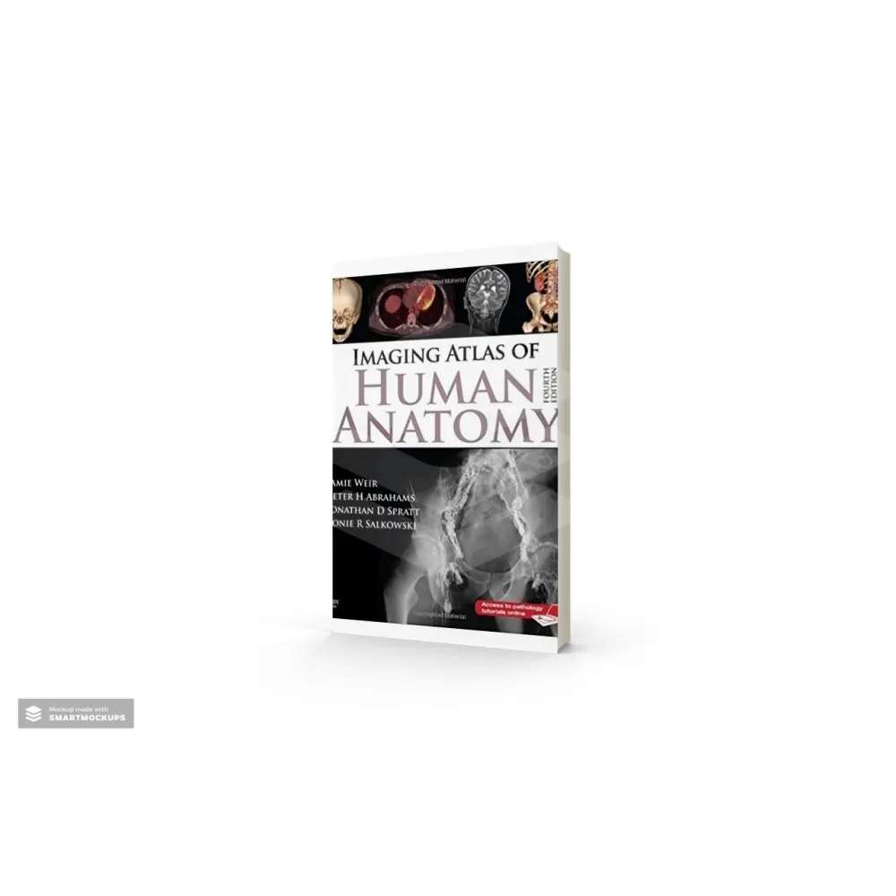
Imaging atlas of Human Anatomy, 5th Edition provides a solid foundation for understanding human anatomy. Jamie Weir, Peter Abrahams, Jonathan D. Spratt, and Lonie Salkowski offer a complete and 3-dimensional view of the structures and relationships within the body through a variety of imaging modalities. Over 60% new images-showing cross-sectional views in CT and MRI, nuclear medicine imaging, and more-along with revised legends and labels ensure that you have the best and most up-to-date visual resource. In addition, you'll get free online access to 10 pathology tutorials linking to additional images for even more complete coverage than ever before (with the opportunity to upgrade to more online tutorials). In print and online, this atlas will widen your applied and clinical knowledge of human anatomy. Imaging is ever more integral to anatomy education and throughout modern medicine. Building on the success of previous editions, this fully revised fifth edition provides a superb foundation for understanding applied human anatomy, offering a complete view of the structures and relationships within the body using the very latest imaging techniques. It is ideally suited to the needs of medical students, as well as radiologists, radiographers and surgeons in training. It will also prove invaluable to the range of other students and professionals who require a clear, accurate, view of anatomy in current practice.
- No. of pages: 280
- Language: English
- Copyright: © Elsevier 2017
Imaging Atlas of Human Anatomy - 5th edition
Imaging atlas of Human Anatomy, 5th Edition provides a solid foundation for understanding human anatomy. Jamie Weir, Peter Abrahams, Jonathan D. Spratt, and Lonie Salkowski offer a complete and 3-dimensional view of the structures and relationships within the body through a variety of imaging modalities. Over 60% new images-showing cross-sectional views in CT and MRI, nuclear medicine imaging, and more-along with revised legends and labels ensure that you have the best and most up-to-date visual resource. In addition, you'll get free online access to 10 pathology tutorials linking to additional images for even more complete coverage than ever before (with the opportunity to upgrade to more online tutorials). In print and online, this atlas will widen your applied and clinical knowledge of human anatomy. Imaging is ever more integral to anatomy education and throughout modern medicine. Building on the success of previous editions, this fully revised fifth edition provides a superb foundation for understanding applied human anatomy, offering a complete view of the structures and relationships within the body using the very latest imaging techniques. It is ideally suited to the needs of medical students, as well as radiologists, radiographers and surgeons in training. It will also prove invaluable to the range of other students and professionals who require a clear, accurate, view of anatomy in current practice.
- No. of pages: 280
- Language: English
- Copyright: © Elsevier 2017
-
- Fully revised legends and labels and over 80% new images – featuring the latest imaging techniques and modalities as seen in clinical practice
-
- Covers the full variety of relevant modern imaging – including cross-sectional views in CT and MRI, angiography, ultrasound, fetal anatomy, plain film anatomy, nuclear medicine imaging and more – with better resolution to ensure the clearest anatomical views
-
- Unique new summaries of the most common, clinically important anatomical variantsfor each body region – reflects the fact that around 20% of human bodies have at least one clinically significant variant
-
- New orientation drawings – to help you understand the different views and the 3D anatomy of 2D images, as well as the conventions between cross-sectional modalities
Now a more compete learning package than ever before, with superb new BONUS electronic enhancements embedded within the accompanying eBook, including:
- Labelled image ‘stacks’ - that allow you to review cross-sectional imaging as if using an imaging workstation
- Labelled image ‘slide-lines’ - showing features in a full range of body radiographs to enhance understanding of anatomy in this essential modality
- Self-test image ‘slideshows’ with multi-tier labelling - to aid learning and cater for beginner to more advanced experience levels
- Labelled ultrasound videos - bring images to life, reflecting this increasingly clinically practiced technique
- Questions and answers accompany each chapter - to test your understanding and aid exam preparation
- 34 pathology tutorials – based around nine key concepts and illustrated with hundreds of additional pathology images, to further develop your memory of anatomical structures and lead you through the essential relationships between normal and abnormal anatomy
- Εκδότης
- Elsevier
- Επιστημονικό πεδίο
- Ανατομία Φυσιολογία
- Επιστημονικό πεδίο
- Μυοσκελετικά
-
- Fully revised legends and labels and over 80% new images – featuring the latest imaging techniques and modalities as seen in clinical practice
-
- Covers the full variety of relevant modern imaging – including cross-sectional views in CT and MRI, angiography, ultrasound, fetal anatomy, plain film anatomy, nuclear medicine imaging and more – with better resolution to ensure the clearest anatomical views
-
- Unique new summaries of the most common, clinically important anatomical variantsfor each body region – reflects the fact that around 20% of human bodies have at least one clinically significant variant
-
- New orientation drawings – to help you understand the different views and the 3D anatomy of 2D images, as well as the conventions between cross-sectional modalities
Now a more compete learning package than ever before, with superb new BONUS electronic enhancements embedded within the accompanying eBook, including:
- Labelled image ‘stacks’ - that allow you to review cross-sectional imaging as if using an imaging workstation
- Labelled image ‘slide-lines’ - showing features in a full range of body radiographs to enhance understanding of anatomy in this essential modality
- Self-test image ‘slideshows’ with multi-tier labelling - to aid learning and cater for beginner to more advanced experience levels
- Labelled ultrasound videos - bring images to life, reflecting this increasingly clinically practiced technique
- Questions and answers accompany each chapter - to test your understanding and aid exam preparation
- 34 pathology tutorials – based around nine key concepts and illustrated with hundreds of additional pathology images, to further develop your memory of anatomical structures and lead you through the essential relationships between normal and abnormal anatomy

Imaging atlas of Human Anatomy, 5th Edition provides a solid foundation for understanding human anatomy. Jamie Weir, Peter Abrahams, Jonathan D. Spratt, and Lonie Salkowski offer a complete and 3-dimensional view of the structures and relationships within the body through a variety of imaging modalities. Over 60% new images-showing cross-sectional views in CT and MRI, nuclear medicine imaging, and more-along with revised legends and labels ensure that you have the best and most up-to-date visual resource. In addition, you'll get free online access to 10 pathology tutorials linking to additional images for even more complete coverage than ever before (with the opportunity to upgrade to more online tutorials). In print and online, this atlas will widen your applied and clinical knowledge of human anatomy. Imaging is ever more integral to anatomy education and throughout modern medicine. Building on the success of previous editions, this fully revised fifth edition provides a superb foundation for understanding applied human anatomy, offering a complete view of the structures and relationships within the body using the very latest imaging techniques. It is ideally suited to the needs of medical students, as well as radiologists, radiographers and surgeons in training. It will also prove invaluable to the range of other students and professionals who require a clear, accurate, view of anatomy in current practice.
- No. of pages: 280
- Language: English
- Copyright: © Elsevier 2017
 ΑΓΟΡΑΣΕ ΚΑΙ ΤΗΛΕΦΩΝΙΚΑ:


 Διαγνωστική απεικόνιση και ψηλάφηση


