

-
-
 ΠΡΟΪΟΝΤΑ
-
-
-
- Ζέρσεϋ
- Επίδεσμοι δακτύλων
- Ελαστικοί Επίδεσμοι
- Tape συγκράτησηs
- Περιποίηση δέρματος και έλκους
- Εργαλεία μέτρησης και θεραπείας
- Ανομοιόμορφα υποστρώματα
- Αφρώδη υποστρώματα και επιθέματα
- Πακέτα Υλικών Θεραπείας
- Αυτοπερίδεση - Περίδεση ύπνου
- Βοηθήματα εφαρμογής ενδυμάτων
- Ενδύματα διαβαθμισμένης συμπίεσης
- Προστατευτικά μπάνιου
-
-
-
-
-
-
-
-
-
-
-
-
-
-
-
-
-
- Μυοσκελετικά
- Θεραπευτική άσκηση
- Εμβιομηχανική - Κινησιολογία
- Manual Therapy
- Κλινικός συλλογισμός
- Θεραπευτικές τεχνικές
- Trigger Points
- Διαχείριση περιτονίας
- Νευρολογικά
- Taping - Περίδεση
- Ανατομία - Φυσιολογία
- Εναλλακτικές θεραπείες
- Βελονισμός
- Αφίσες ανατομίας
- Λεξικά
- Οίδημα - Λεμφοίδημα
- Ψυχολογία
- Οστεοπαθητική
- Aναπνευστική/Καρδιαγγειακή φυσικοθεραπεία
- Αθλητική φυσικοθεραπεία
- Προσφορές
-
-
-
-
-
-
-
-
-
-
-
-
-
-
 ESSENTIALS
-
 SUPER OFFERS
-
 ΕΠΙΚΟΙΝΩΝΙΑ
-
 BLOG
BLOG
-
 BRANDS
-

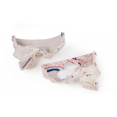
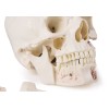
This skull model is an actual cast of a real human specimen and shows all anatomical structures in the highest detail. It is made for students in anatomy, medicine, surgery, otolaryngology, ophthalmology, and dentistry. The Skull is intricately sectioned and reassembled with metal and magnet connections.
Deluxe demonstration skull - 14 parts for advanced studies
This skull model is an actual cast of a real human specimen and shows all anatomical structures in the highest detail. It is made for students in anatomy, medicine, surgery, otolaryngology, ophthalmology, and dentistry. The Skull is intricately sectioned and reassembled with metal and magnet connections.
This skull model is an actual cast of a real human specimen and shows all anatomical structures in the highest detail. It is made for students in anatomy, medicine, surgery, otolaryngology, ophthalmology, and dentistry. The Skull is intricately sectioned and reassembled with metal and magnet connections.
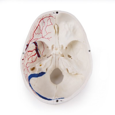
The calvarium is sectioned horizontally leaving the temporal bone and its sutures intact. Bony impressions of the superior sagittal sinus, transverse sinus, and sigmoid sinus as well as the meningeal vessels have been painted. The base portion of the skull has been sagittally sectioned in the way that it passes through the one cribriform plate on one side and another section in the same plane passes through the other cribriform plate of the ethmoid leaving the crista Galli perpendicular plate of the ethmoid intact as also the whole nasal septum.
The structures of the anterior, middle, and posterior cranial fossae are easily accessible. One can directly visualize the nasal cavity, the concha, the nasal septum, and the bony pharyngeal and nasopharyngeal spaces. The nasal septum is separable from surrounding bones. The frontal sinuses have been dissected on one side to show the sinus as a whole and on the other side chiseled out for full access to the sinus. The relation of this sinus to the nasal cavity is clearly shown and is especially valuable for otolaryngologists.
On one side of the skull, the temporal bone has been left in situ. The other temporal bone is removable from the skull. A portion of the mastoid and squama can be removed along with the tympanic antrum, bearing the internal ear in full view. All three semicircular canals are visible along with the course of the facial nerve coursing backward and then downwards emerging finally through the stylo-mastoid foramen.
The removable temporal bone has the external auditory meatus intact. An almost vertical section through the squama mastoid process and carried inwards along the petro-squamosal junction has been made and when apart, one sees the position of the tympanic membrane. The carotid canal has been opened as also the cochlea, showing the internal canal, and the course of the facial nerve has been depicted. The oval window, the semi-circular canals, and aditus of the tympanic antrum are visible.
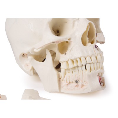
The maxilla and mandible expose the structures of dentition, and the roots, the bony margin of the alveolar process, dental vessels, and nerves are visible. The maxillary sinus can be opened by removing a bone flap. The teeth of the right mandible are sectioned to show the inner tooth structure.

This skull model is an actual cast of a real human specimen and shows all anatomical structures in the highest detail. It is made for students in anatomy, medicine, surgery, otolaryngology, ophthalmology, and dentistry. The Skull is intricately sectioned and reassembled with metal and magnet connections.
 ΑΓΟΡΑΣΕ ΚΑΙ ΤΗΛΕΦΩΝΙΚΑ :








 ΠΡΟΠΛΑΣΜΑΤΑ ΑΝΑΤΟΜΙΑΣ


